Rosacea
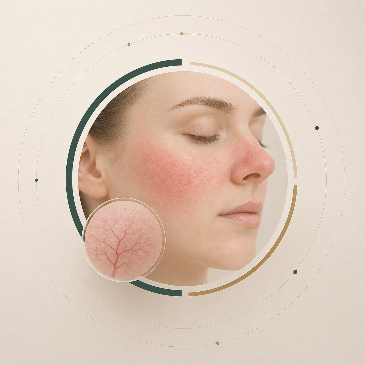
Chronic facial redness, flushing and visible vessels. Management includes trigger avoidance, topical and oral therapies, and light-based treatments where appropriate. Individual plan depends on subtype and severity.
Medical skin consultation — doctor-led assessment, individual treatment suitability review.
LP Clinic offers medical dermatology consultations alongside its hair restoration services. The approach is diagnosis-led — a qualified specialist assesses the skin condition, confirms what treatment (if any) is appropriate, and develops an individual plan.
This page describes the consultation approach in general terms. The clinic team responds individually and explains which services are suitable for your case.

You describe your concern — via WhatsApp or in person. Photographs may be shared (only after privacy consent). The medical team reviews the information before responding.
A qualified specialist evaluates the skin condition. This may include visual examination, medical history review, and where appropriate, diagnostic tests.
If treatment is appropriate, options are discussed. The range of available treatments and their suitability for your specific case are assessed individually.
Where treatment is recommended, an individual plan is developed. No treatment is initiated without your understanding and consent.
A reference of conditions frequently assessed at LP Clinic. Every case requires individual examination — the notes below describe the general approach, not a diagnosis or treatment recommendation.
Chronic facial redness, flushing and visible vessels. Management includes trigger avoidance, topical and oral therapies, and light-based treatments where appropriate. Individual plan depends on subtype and severity.
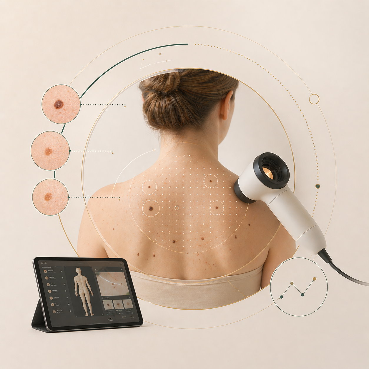
Digital dermoscopic mapping of pigmented skin lesions for early detection of change. Baseline imaging is taken and compared over time. Recommended for patients with numerous moles, previous skin cancer, or family history.

Chronic inflammatory skin condition with characteristic plaques. Treatment is tailored to extent and location — topicals, phototherapy, and systemic options all have a role. Long-term management planning is important.
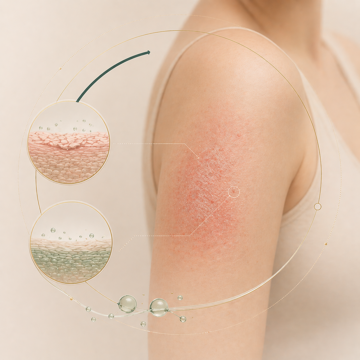
Chronic eczema with dry, itchy, inflamed skin. Treatment combines skin barrier repair, topical anti-inflammatory therapy and trigger management. May be assessed alongside allergology where relevant.
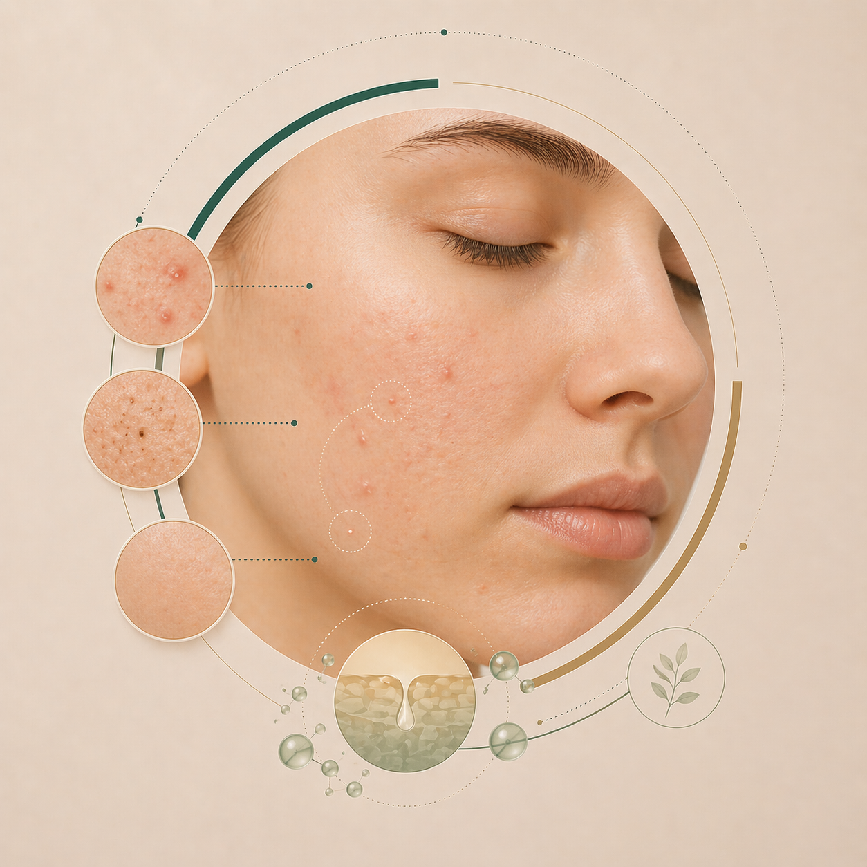
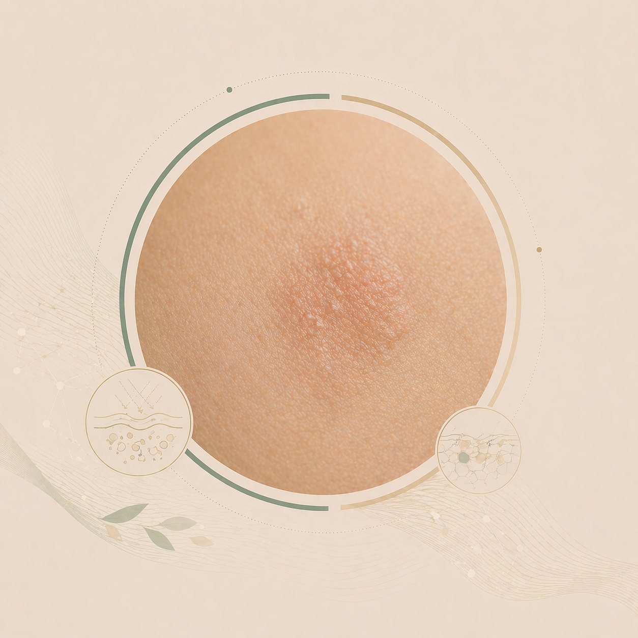
Inflammatory skin condition with comedones, papules and sometimes cysts, most often on the face. Treatment ranges from topicals to oral therapies and adjunctive procedures — tailored to severity and skin type.
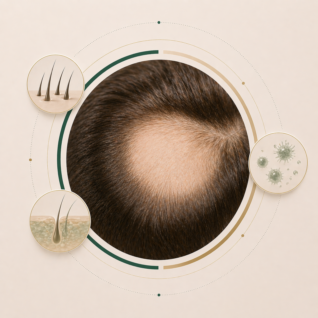
Autoimmune form of hair loss presenting as well-defined, round patches. Assessment clarifies the pattern and possible triggers; treatment is individualised and accounts for the often relapsing course.
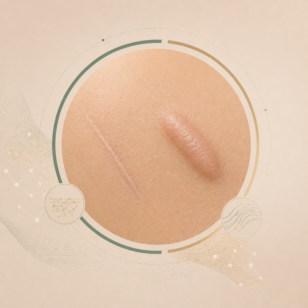
Abnormal scar formation following injury, surgery or inflammation. Treatment ranges from intralesional injections and light-based procedures to surgical revision — depending on scar type, location and age.
UV-induced skin change and early form of skin cancer. Assessment includes dermoscopy and biopsy where indicated; treatment is stage-adapted — from topicals and cryotherapy to physical procedures.
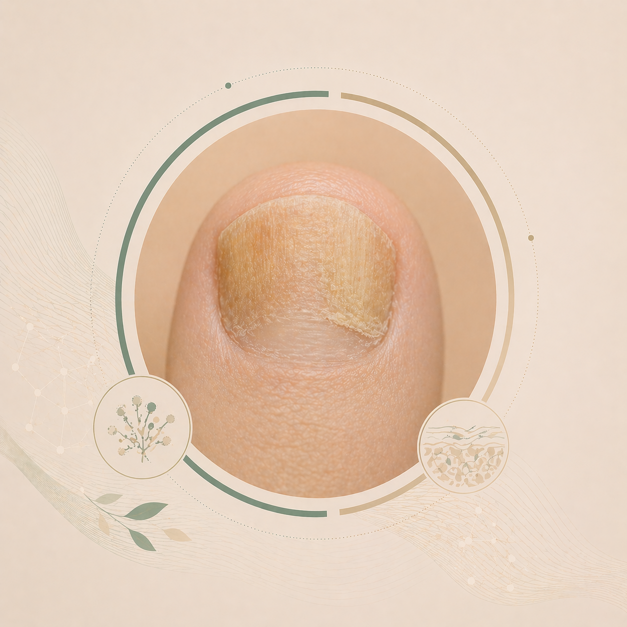
Fungal infection of finger- or toenails with discoloration, thickening and brittleness. Diagnosis is confirmed by laboratory testing; treatment combines topical and, where needed, systemic therapy over several months.
LP Clinic also assesses other conditions including mole removal, hyperpigmentation, seborrheic hyperkeratosis, warts, herpes zoster, and age-related skin changes. Contact the clinic to ask whether an appointment is appropriate for your concern.
This page does not provide a medical diagnosis, treatment recommendation, or prescription. Skin conditions require individual clinical evaluation by a qualified specialist. Do not use this information to self-diagnose or self-treat. If you have an urgent skin concern, consult a medical professional promptly.